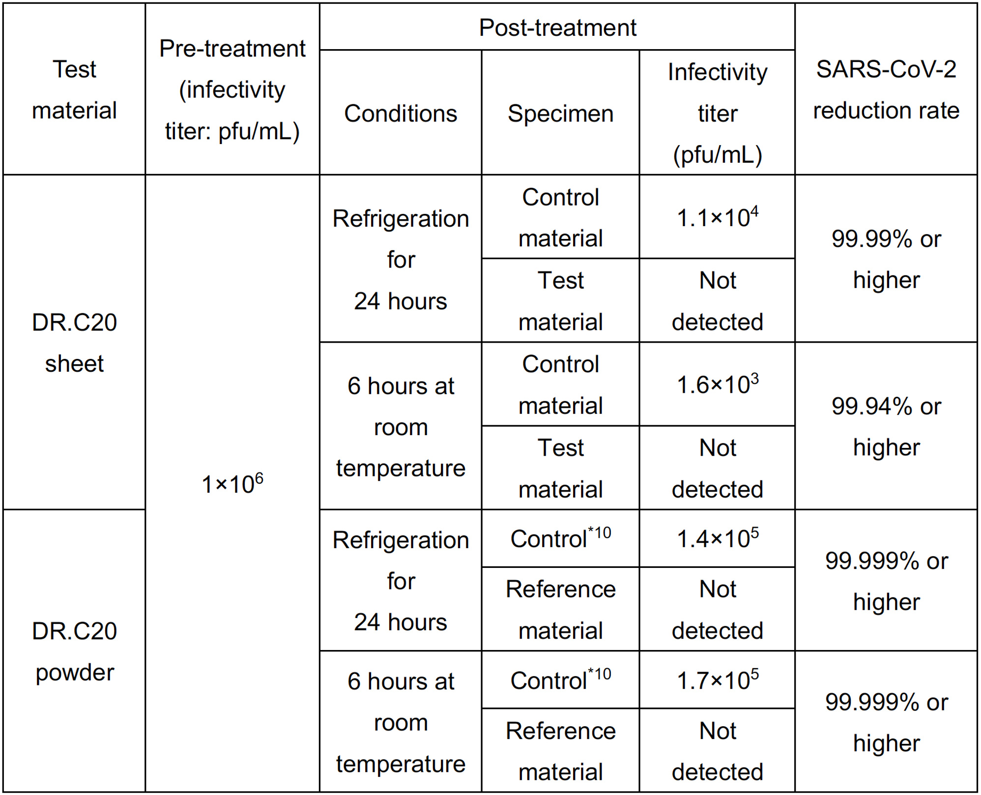
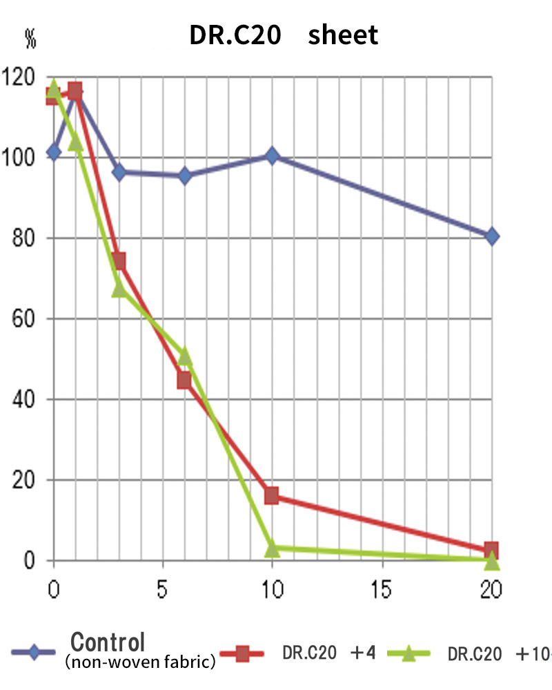
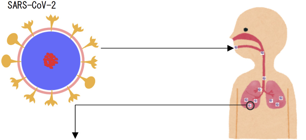
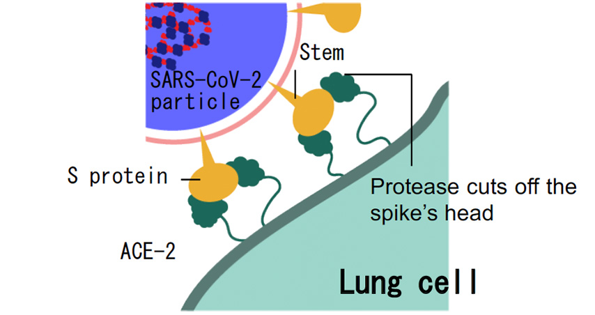
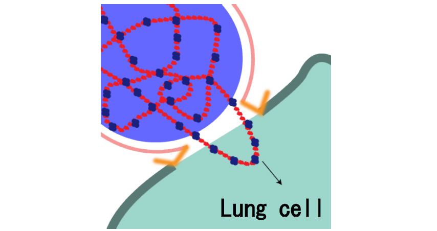
Research on SARS-CoV-2 spike proteins disintegration by DR.C20

Janualy 13, 2021
DR.C Medical Medicine Co., Ltd.

1 DR.C Medical Medicine has verified that the SARS-CoV-2*1 infectivity titer*2 is reduced by 99.99% using a sheet*3 adhered with photocatalytic DR.C20*4 powder.(Joint research with Nagasaki University)

2 World-first*5 – We have verified through biochemical analysis that DR.C20 cuts artificially-synthesized SARS-CoV-2 spike proteins*6 in several locations in a period between 3 and 10 minutes. A molecular dynamics simulation suggested that such cutting action breaks down the 3D structure of the spike protein necessary for it to bond with the human cell receptor, ACE2*7 .(Joint research with Institute of Physical and Chemical Research)

Overview

DR.C Medical Medicine Co., Ltd. (Representative Director: Narumi Okazaki), in a joint initiative with Professor Jiro Yasuda of the world-renown infectious disease institute, National Research Center for the Control and Prevention of Infectious Diseases Nagasaki University (CCPID) (also a Professor of Emerging Infectious Diseases at the Institute of Tropical Medicine) has confirmed that the SARS-CoV-2 infectivity titer was reduced by 99.99% when a sheet adhered with DR.C20 powder (hereinafter “DR.C20 sheet”) was immersed in SARS-CoV-2 solution and estimates that the infectivity titer will decrease by 99% or more within 5 minutes

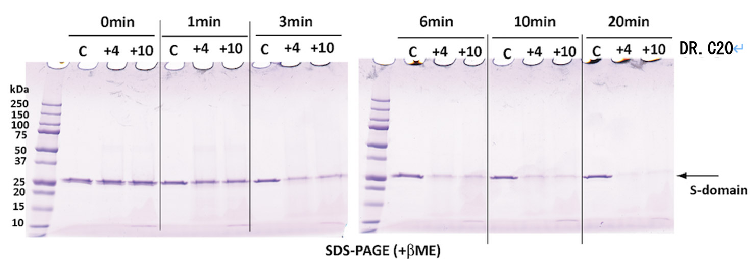
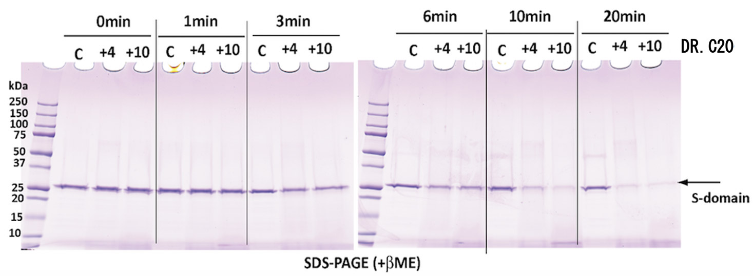
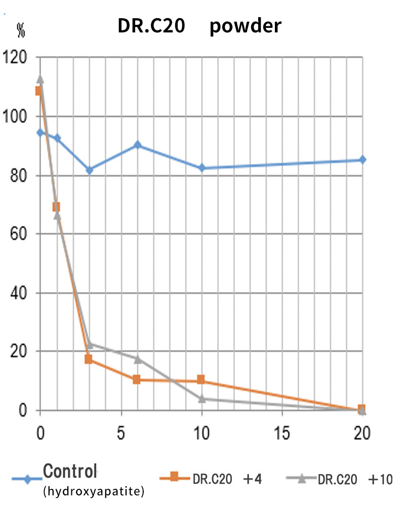
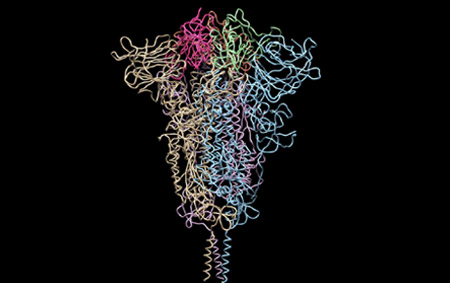
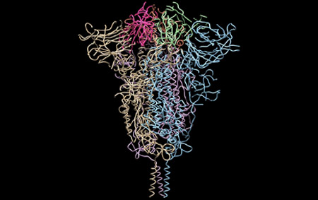
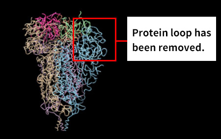
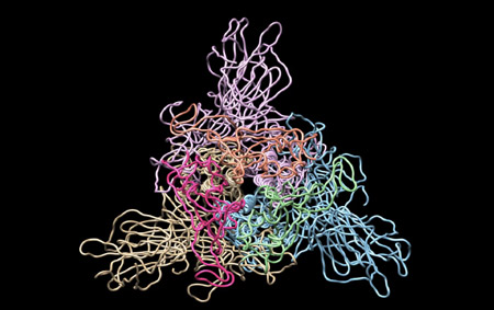
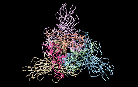
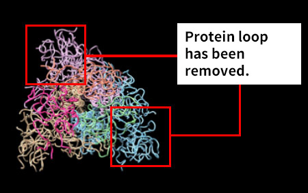
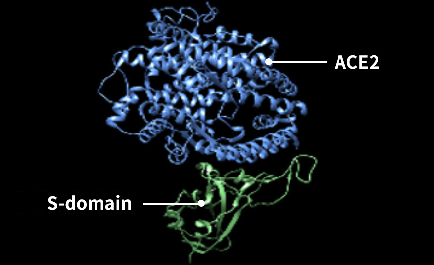
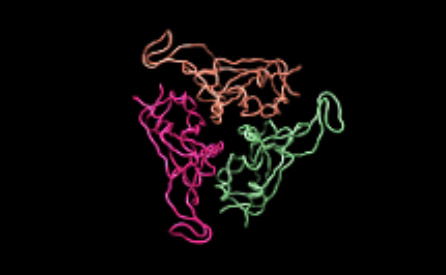
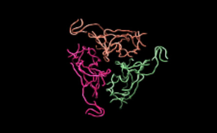
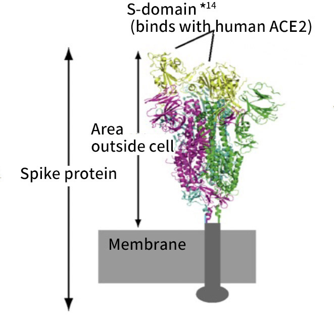
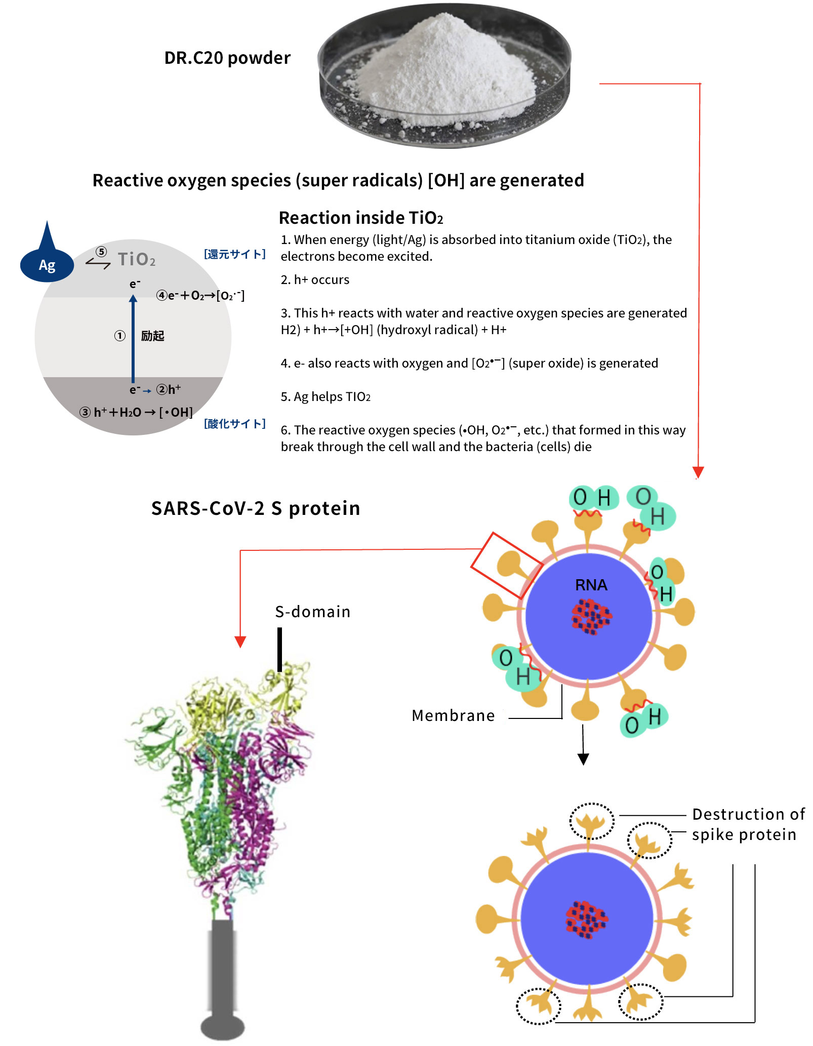
Moreover, together with Visiting Professor Shigeyuki Yokoyama of RIKEN’s Yokoyama Laboratory, an institution internationally renowned for protein synthesis, as a result of conducting an experiment using artificially-synthesized spike protein related to SARS-CoV-2 infection, we verified that DR.C20 cut the protein in multiple locations within a period of 3 to 10 minutes. The result of a molecular dynamics simulation carried out with the support of Associate Professor Toru Terada at the Graduate School of Agricultural and Life Sciences, The University of Tokyo, suggested that such cutting action by DR.C20 breaks down the 3D structure of the spike protein necessary for it to bond with the human cell receptor, ACE2. We clarified that these effects of DR.C20 on spike protein led to a reduction in the virus infectivity titer.

Background

COVID-19, which broke out in December 2019, is an extremely infectious virus that has resulted in 86.47 million infected persons and over 1.87 million deaths*8 throughout the world as of January 2021. There are demands for immediate measures in various fields to address this urgent social issue.

This time, in addition to verifying a reduction in SARS-CoV-2 infectivity titer jointly with Nagasaki University, we also conducted joint research with Visiting Professor Shigeyuki Yokoyama of RIKEN’s Yokoyama Laboratory to confirm the details of the breakdown mechanism of SARS-CoV-2 proteins.
Human cells have a molecular structure enabling SARS-CoV-2 to efficiently penetrate them. The ACE2 receptor, used as the “doorway” to a cell, comprises of the proteases, TMPRSS2 and/or FURIN. SARS-CoV-2 first binds its spike protein (Spike, S) protruding on the surface with the host cell’s ACE2 receptor. The proteases TMPRSS2 and FURIN inside the cell membrane are known to cut the virus’s spike protein at an appropriate position and help the virus and the cell to fuse. Therefore, it is believed that if the SARS-CoV-2 spike protein is stopped from functioning, it would no longer be possible for SARS-CoV-2 to infect human cells.

We are reporting the results of this research as we believe they can contribute to society as a COVID-19 infection prevention measure.

*1 Severe acute respiratory syndrome coronavirus 2 (SARS-CoV-2). The virus responsible for coronavirus disease 2019 (COVID-19).
*2 The viral load with infectivity.
*3 Polyester fiber
*4 About DR.C20 technology
DR.C20, a new titanium oxide compound material was developed by DR.C Medical Medicine leveraging original technology by applying photocatalysis. The titanium oxide material generates an OH (hydroxyl) radical with extremely high oxidizability from the surrounding water to break down the protein on the virus’s surface through chemical reaction.
*5 According to an investigation by DR.C Medical Medicine, current as of Jan.7.2021.
*6 A type of protein appearing on the surface of SARS-CoV-2. There is also envelope protein ((E); important in viral infection) and matrix protein ((M); interacts with E protein). It is believed that the virus penetrates the cell when this spike protein bonds with the receptor of the human cells.
*7 An abbreviation for Angiotensin-Converting Enzyme 2. The receptor used as the “doorway” to human cells.
*8 According to a count by Johns Hopkins University, U.S.A. (current as of Jan.6.2021)

Experiment Description① (Institute of Tropical Medicine, Nagasaki University)

  • ◆Performing institution:
    Institute of Tropical Medicine/Emerging Infectious Diseases, Nagasaki University
  • ◆Aim:
    Measure the effect over time of DR.C20 reducing the infectivity titer of SARS-CoV-2.
  • ◆Method:
    Immersed a DR.C sheet in a virus solution (1×10⁶pfu/mL), collected virus solution after allowing to stand for a set period of time, then measured the viral load (virus infectivity titer according to a plaque assay*9) (Performed in the BSL-3 laboratory)

*9 Plaque assay is a method using the number of cells that form cell lysates (plaque) when virus-infected cells denature or are destroyed as an indicator of virus load.

Fig. 1 A DR.C20 sheet immersed in virus solution

Fig. 2 Cell (Vero E6) used to measure virus infectivity titer

  • ◆Results:

Table 1. Antiviral activity of DR.C20 against SARS-CoV-2

*10 Untreated group (SARS-CoV-2 virus solution only)

Fig. 3 Image of plaque in SARS-CoV-2 infectivity titer test(photo of plaque assay result on specimen refrigerated for 24 hours)

Photo 1 (Control material: Sh-NC): When the cultured cells are infected by SARS-CoV-2, they are destroyed, therefore appear white and transparent. This indicates the existence of SARS-CoV-2 that has not been inactivated.

Photo 2 (Test material: SH+10):The SARS-CoV-2 solution is not white and transparent. This indicates that the virus has been inactivated and the cells have not been destroyed.

Fig. 4 Virus decay rate

The results of an analysis using the above virus decay curve confirmed that sheet adhered with DR.C20 powder reduced virus infectivity titer by 99% in 5 minutes.

◆Conclusion from experiment at Nagasaki University

The experiment verified that SARS-CoV-2 infectivity titer decreased by 99.99% when a sheet adhered with DR.C20 powder was immersed in SARS-CoV-2 solution, and it is estimated that the infectivity titer will decrease by 99% or more within 5 minutes.

Experiment Description② (RIKEN)

  • ◆Performing institution:
    RIKEN’s Yokoyama Laboratory
  • ◆Aim:
    Yokoyama Laboratory will artificially synthesize and adjust a SARS-CoV-2 spike protein (S)*11 sample and analyze how that structure is impaired by DR.C20. A molecular dynamics calculation will then be used to analyze the impact of this impairment on the 3D structure of spike protein.
  • ◆Method:
    DR.C20 powder (two types; low concentration titanium +4 and low concentration titanium +10) an adhered sheet, and SARS-CoV-2 protein will be treated under each condition, then treated samples of the control group and test group will be analyzed with SDS-PAGE*12.
  • ◆Molecular dynamics calculation*13:
    In regards to the spike protein, for which the 3D structure is already known, with the support of Associate Professor Tohru Terada majoring in Applied Life Science at the Graduate School of Agricultural and Life Sciences (BINDS), utilize molecular dynamics simulation, in other words calculate molecular movement with a computer, in order to visualize the process of cutting spike protein and breaking down the 3D structure of its modified proteins. spike proteins are extremely large, therefore a coarse grained model will be used which treats around four atoms (excluding hydrogen atoms) as one particle.

*11 A spike protein exists in a form whereby it pierces the lipid bilayer that makes up a virion’s surface.
*12 SDS-polyacrylamide gel electrophoresis. In order to obtain the results of phoresis only reflecting the protein’s peptide chain length, electrophoresis utilizes the surfactant with a negative charge, SDS (sodium dodecyl sulfate = sodium lauryl sulfate) and polyacrylamide gel. The sample is denatured using SDS, cancelling the charge specific to the protein to achieve uniformity, therefore results can be used in the estimation of molecular weight.
*13 Shows the simulated molecule movement based on measurement results.

  • ◆Results

(1)Electrophoresis SDS-PAGE (+βME)

① If the S-domain sample solution is illuminated with artificial sunlight in the presence of DR.C20 (powder sample), we believe that cutting will begin after 3 minutes.

C:Control (no DR.C20)
+4: Low concentration DR.C20    +10: Low concentration DR.C20

②If the S-domain sample solution is illuminated with artificial sunlight in the presence of DR.C20 (sheet sample), we believe that cutting on the DR.C20 sheet sample will begin after 3 minutes.

C: Control (no DR.C20)
+4: Low concentration DR.C20   +10: Low concentration DR.C20

(2)If the S-domain sample solution is illuminated with artificial sunlight in the presence of DR.C20, it will be cut and destroyed in a period of 3 to 10 minutes.
Percentage over time of protein quantity destroyed compared to the control.

◆In the case of the DR.C20 powder sample, cutting begins after 1 minute, and at least 80% of the S-domain has been destroyed after 6 minutes.

◆In the case of the DR.C20 sheet sample, cutting begins after 3 minutes, and at least 80% of the S-domain has been destroyed after 10 minutes.

(3) Molecular dynamics (MD) simulation of the SARS-CoV-2 spike protein

①A video of a molecular dynamics simulation showing that the destruction of the 3D structure of a cut spike protein was triggered practically instantly (2 microseconds: 2 x 10⁻⁶ seconds). The below figures are side views.

Native state(when stable)

The spike protein trimer in a native state is stable and the 3D structure is preserved.

Native state
(when stable)

The spike protein trimer in a native state is stable and the 3D structure is preserved.

(Cutting)

If the protein loop is cut, the 3D structure deteriorates instantly and destruction begins.

(After break down)

The 3D structure swiftly deteriorates and is destroyed.

②A video of a molecular dynamics simulation showing that the destruction of the 3D structure of a cut spike protein was triggered practically instantly (2 microseconds: 2 x 10⁻⁶ seconds). These figures are top (external) views.

Native state(when stable)

The spike protein trimer in a native state is stable and the 3D structure is preserved.

Native state
(when stable)

The spike protein trimer in a native state is stable and the 3D structure is preserved.

(Cutting)

If the protein loop is cut, the 3D structure deteriorates instantly and destruction begins.

The 3D structure swiftly deteriorates and is destroyed.

(4)Molecular dynamics (MD) simulation focusing on the spike protein S-domain and ACE2 receptor

RBD and ACE2 compound (PDB ID: 6M0J)

The S-domain in a native state is stable and the 3D structure is preserved.

RBD and ACE2 compound
(PDB ID: 6M0J)

The S-domain in a native state is stable and the 3D structure is preserved.

(Cutting)

If the protein loop is cut, the 3D structure deteriorates instantly and destruction begins.

The 3D structure swiftly deteriorates and is destroyed.

SARS-CoV-2 spike protein trimer

1   Analysis of breakdown/cutting with DR.C20

If the solution containing the artificially-synthesized S-domain sample is illuminated with artificial sunlight in the presence of DR.C20, it will be cut in a period of 3 to 10 minutes.

2   Analysis of impact of cutting on 3D structure

A molecular dynamics (MD) simulation showed that the cutting of the SARS-CoV-2 spike protein by DR.C20 swiftly triggers deterioration/destruction of the 3D structure. This renders the SARS-CoV-2 virus incapable of binding with ACE2.

*14 S-domain: The portion within the SARS-CoV-2 spike protein particularly important for binding with human cell receptors.

Conclusion from experiment at RIKEN   Cutting by DR.C20 swiftly triggers deterioration/destruction of the 3D structure

S-domain can no longer bind with ACE2

We have verified through biochemical analysis that DR.C20 cuts artificially-synthesized SARS-CoV-2 spike protein*6 in several locations in a period between 3 and 10 minutes. A molecular dynamics simulation suggested that such cutting action breaks down the 3D structure of the spike protein necessary for it to bond with the human cell receptor, ACE2*7.

To date, using a cell-free protein synthesis, we have also succeeded in manufacturing envelope protein (E) and matrix protein (M) and plan to carry out biochemical analysis applying the same method as spike protein.

■ Summary from joint research by Nagasaki University and RIKEN

The results obtained by Professor Yasuda of the National Research Center for the Control and Prevention of Infectious Diseases Nagasaki University (CCPID), confirmed that the SARS-CoV-2 infectivity titer*2 was reduced by 99.99% and estimated that the infectivity titer will decrease by 99% or more within 5 minutes.
The results obtained by Visiting Professor Shigeyuki Yokoyama at RIKEN’s Yokoyama Laboratory suggested that DR.C20 cut and destroyed the artificially-synthesized SARS-CoV-2 spike protein in a period of 3 to 10 minutes and destroyed the 3D structure necessary for the spike protein to bind with the human cell receptor, ACE2. In light of the above results, we believe that by applying DR.C20 powder and sheets to various goods, we can increase the extent to which these findings contribute to society.

《Mechanism of SARS-CoV-2 penetrating human lung cells (Illustration 1)》

Binding with lung cell

After the virus’s spike protein binds with the ACE2 receptor, the lung cell’s proteases (enzymes catalyzing breakdown of proteins) cut the head of the spike off and trigger the fusion mechanism for the virus to enter the cell.

Penetrates inside lung cell

The virus and the membrane of lung cells bind, then the virus’s RNA centers the cell. This RNA is virus genome and contains the genetic code.

《The Proteolysis Mechanism of DR.C20 (Illustration 2)》